2015/12/18
Researchers have designed an optical funnel that could focus a stream of proteins or viruses precisely into the fine beam of an X-ray laser for analysis. The funnel consists of a specially shaped light beam that traps particles at its centre. In contrast to conventional optical traps, the funnel uses thermal forces to control the particles. The team led by Andrei Rode from The Australian National University in Canberra, and including researchers from the Center for Free-Electron Laser Science (CFEL) at DESY and the Center for Ultrafast Imaging (CUI) at the University of Hamburg, tested their concept with micrometre-sized glass and carbon beads, as they report in the scientific journal Physical Review Applied. CFEL is a cooperation of DESY, the University of Hamburg and the Max Planck Society.
Scientists use X-rays to analyse the spatial structure of biomolecules in order to reveal much about their modes of action. This not only leads to a better understanding of biological processes, it can also pave the way for the development of novel drugs. For the analysis, tiny crystals grown from the biomolecules have to be fed into the X-ray beam. The crystals diffract the X-rays in a characteristic way, and from this diffraction pattern the structure of the molecule can be calculated. “As biomolecules are notoriously hard to crystallise, we are also working on ways to analyse single molecules directly, with the power of ultra bright X-ray free-electron lasers or XFELs,” explains co-author Jochen Küpper from DESY, head of the Controlled Molecule Imaging group at CFEL.
In either case, thousands of diffraction images are needed to calculate the molecule's structure. As each crystal or molecule is instantly destroyed by the intense flash from an X-ray laser, many thousand samples have to be streamed across the beam. Usually, just a small fraction of samples in this stream is actually hit by one of the ultra short XFEL flashes. The better the stream of samples is focussed, the more efficient is its use. Current techniques can squeeze particle streams down to about 50 micrometres – but a typical X-ray laser beam with a diameter of 100 nanometres is still 500 times smaller. Methods using liquid jets as sample carriers do better, but the liquid interferes with the diffraction image. “That’s why the optical funnel is so appealing,” says co-author Henry Chapman from DESY, head of the Coherent Imaging Division at CFEL. The trapping force works in air and vacuum, and the small end of the funnel, where particles are directed, is only a few micrometres wide.
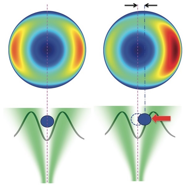
Optical trapping, such as with optical tweezers, typically relies on direct light forces like momentum transfer or electric field effects to manipulate a target object immersed in water. Thermal effects are minimized because the water carries heat away. However, in a gas, the lower thermal conductivity of the surrounding medium allows the object to heat up on the side where the light shines, while remaining cool on the opposite side. Gas molecules collide with the hot side and bounce off with more momentum than those hitting the cold side. This momentum imbalance results in a so-called photophoretic force that pushes from hot to cold, drawing the object toward regions of lower light intensity.
To create their funnel, the scientists used an optical vortex, which is a light beam with a helical wavefront. This results in a dark central region in the beam where the light intensity drops down to zero. By passing the vortex beam through a lens, the physicists gave it a funnel like shape. To demonstrate the concept, they put small graphite beads with 12 to 20 micrometres diameter and some larger graphite coated glass beads into their optical funnel and measured the photophoretic force that acted on the beads. The advantage is that this force can be much stronger than direct optical forces; moreover, the particle spends most time in the dark core of the light trap. Although the measurements do not directly apply to proteins and other biomolecules, the scientists estimate that these will behave similar as they typically have an even smaller thermal conductivity and, thus, will experience a comparable temperature difference. In further experiments, the researchers hope to verify this and to optimise the optical funnel for use in an XFEL.
Reference:
“Optically Induced Forces Imposed in an Optical Funnel on a Stream of Particles in Air or Vacuum”; Niko Eckerskorn, Richard Bowman, Richard A. Kirian, Salah Awel, Max Wiedorn, Jochen Küpper, Miles J. Padgett, Henry N. Chapman, and Andrei V. Rode; Physical Review Applied 4, 064001, 2015; DOI: 10.1103/PhysRevApplied.4.064001 - Selected as Editor’s Choice
Further reading:
American Physical Society „Focus“: https://physics.aps.org/articles/v8/121

