July 24th, 2013
At EPFL’s Blue Brain facilities, computer models of individual neurons are being assembled into neural circuits that produce electrical signals akin to brain waves. The results, published in the journal Neuron, are helping solve the mystery of how and why these signals arise in the brain.
For almost a century, scientists have been studying brain waves to learn about mental health and the way we think. Yet the way billions of interconnected neurons work together to produce brain waves remains unknown. Now, scientists from EPFL’s Blue Brain Project in Switzerland, at the core of the European Human Brain Project, and the Allen Institute for Brain Science in the United States, show in the July 24th edition of the journal Neuron how a complex computer model is providing a new tool to solve the mystery.
The brain is composed of many different types of neurons, each of which carry electrical signals. Electrodes placed on the head or directly in brain tissue allow scientists to monitor the cumulative effect of this electrical activity, called electroencephalography (EEG) signals. But what is it about the structure and function of each and every neuron, and the way they network together, that give rise to these electrical signals measured in a mammalian brain?
Modeling Brain Circuitry
The Blue Brain Project is working to model a complete human brain. For the moment, Blue Brain scientists study rodent brain tissue and characterize different types of neurons to excruciating detail, recording their electrical properties, shapes, sizes, and how they connect.
To answer the question of brain-wave origin, researchers at EPFL’s Blue Brain Project and the Allen Institute joined forces with the help of the Blue Brain modeling facilities. Their work is based on a computer model of a neural circuit the likes of which have never been seen before, encompassing an unprecedented amount of detail and simulating 12,000 neurons.
“It is the first time that a model of this complexity has been used to study the underlying properties of brain waves,” says EPFL scientist Sean Hill.
In observing their model, the researchers noticed that the electrical activity swirling through the entire system was reminiscent of brain waves measured in rodents. Because the computer model uses an overwhelming amount of physical, chemical and biological data, the supercomputer simulation allows scientists to analyze brain waves at a level of detail simply unattainable with traditional monitoring of live brain tissue.
“We need a computer model because it is impossible to relate the electrical activity of potentially billions of individual neurons and the resulting brain waves at the same time,” says Hill. “Through this view, we’re able to provide an interpretation, at the single-neuron level, of brain waves that are measured when tissue is actually probed in the lab.”

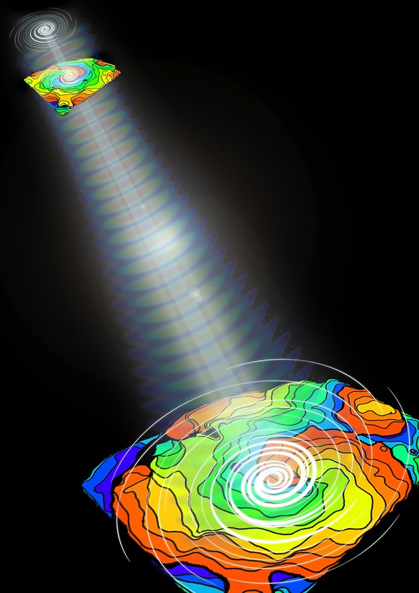
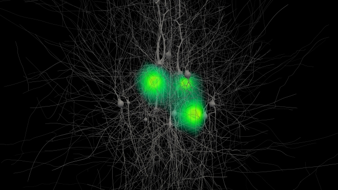
Computer visualization of brain waves produced by 100 detailed models of nerve cells. The strength of the signal is represented by the following color scheme: green = weak signal, yellow = medium, red = strong.
(c) Copyright: EPFL, Blue Brain Project Based on research published on July 24th 2013 in the journal NEURON.
Finding brain wave analogs
Neurons are somewhat like tiny batteries, needing to be charged in order to fire off an electrical impulse known as a “spike”. It is through these “spikes” that neurons communicate with each other to produce thought and perception. To “recharge” a neuron, charged particles called ions must travel through miniscule ionic channels. These channels are like gates that regulate electrical current. Ultimately, the accumulation of multiple electrical signals throughout the entire circuit of neurons produces brain waves.
The challenge for scientists in this study was to incorporate into the simulation the thousands of parameters, per neuron, that describe these electrical properties. Once they did that, they saw that the overall electrical activity in their model of 12,000 neurons was akin to observations of brain activity in rodents, hinting at the origin of brain waves.
“Our model is still incomplete, but the electrical signals produced by the computer simulation and what was actually measured in the rat brain have some striking similarities,” says Allen Institute scientist Costas Anastassiou.
Hill adds, “For the first time, we show that the complex behavior of ion channels on the branches of the neurons contributes to the shape of brain waves.”
There is still much work to be done in order to arrive at a complete simulation. While the model’s electrical signals are analogous to in vivo measurements, researchers warn that there are still many open questions as well as room to improve the model. For instance, the simulation is modeled on neurons that control the hind-limb, while in vivo data represent brain waves coming from neurons that have a similar function but control whiskers instead.
“Even so, the computer model we used allowed us to characterize, and more importantly quantify, key features of how neurons produce these signals,” says Anastassiou.
The scientists are currently studying similar brain wave phenomena in larger and more realistic neural circuits.
This computer model is drawing cellular biophysics and cognitive neuroscience closer together, in order to achieve the same goal: understanding the brain. But the two disciplines share neither the methods nor the scientific language. By simulating electrical brain activity and relating the behavior of single neurons to brain waves, the researchers aim to bridge this gap, opening the way to better tools for diagnosing mental disorders, and on a deeper level, offering a better understanding of ourselves.
About the Allen Institute for Brain Science
The Allen Institute for Brain Science (www.alleninstitute.org) is an independent, 501(c)(3) nonprofit medical research organization dedicated to accelerating the understanding of how the human brain works in health and disease. Using a big science approach, the Allen Institute generates useful public resources used by researchers and organizations around the globe, drives technological and analytical advances, and discovers fundamental brain properties through integration of experiments, modeling and theory. Launched in 2003 with a seed contribution from founder and philanthropist Paul G. Allen, the Allen Institute is supported by a diversity of government, foundation and private funds to enable its projects. Given the Institute’s achievements, Mr. Allen committed an additional $300 million in 2012 for the first four years of a ten-year plan to further propel and expand the Institute’s scientific programs, bringing his total commitment to date to $500 million. The Allen Institute’s data and tools are publicly available online at www.brain-map.org.
About EPFL
With over 350 laboratories and research groups on campus, EPFL in Lausanne, Switzerland, is one of Europe’s most innovative and productive scientific institutions. Ranked top 3 in Europe and top 20 worldwide in many scientific rankings, EPFL has attracted the best researchers in their fields. The institute’s unique structure fosters transdisciplinary research and promotes partnerships with other institutions. EPFL at a glance : http://information.epfl.ch/glance Latest scientific news : http://news.epfl.ch