February 16, 2016
University of Colorado Boulder researchers have demonstrated the use of the world’s first ultrafast optical microscope, allowing them to probe and visualize matter at the atomic level with mind-bending speed.
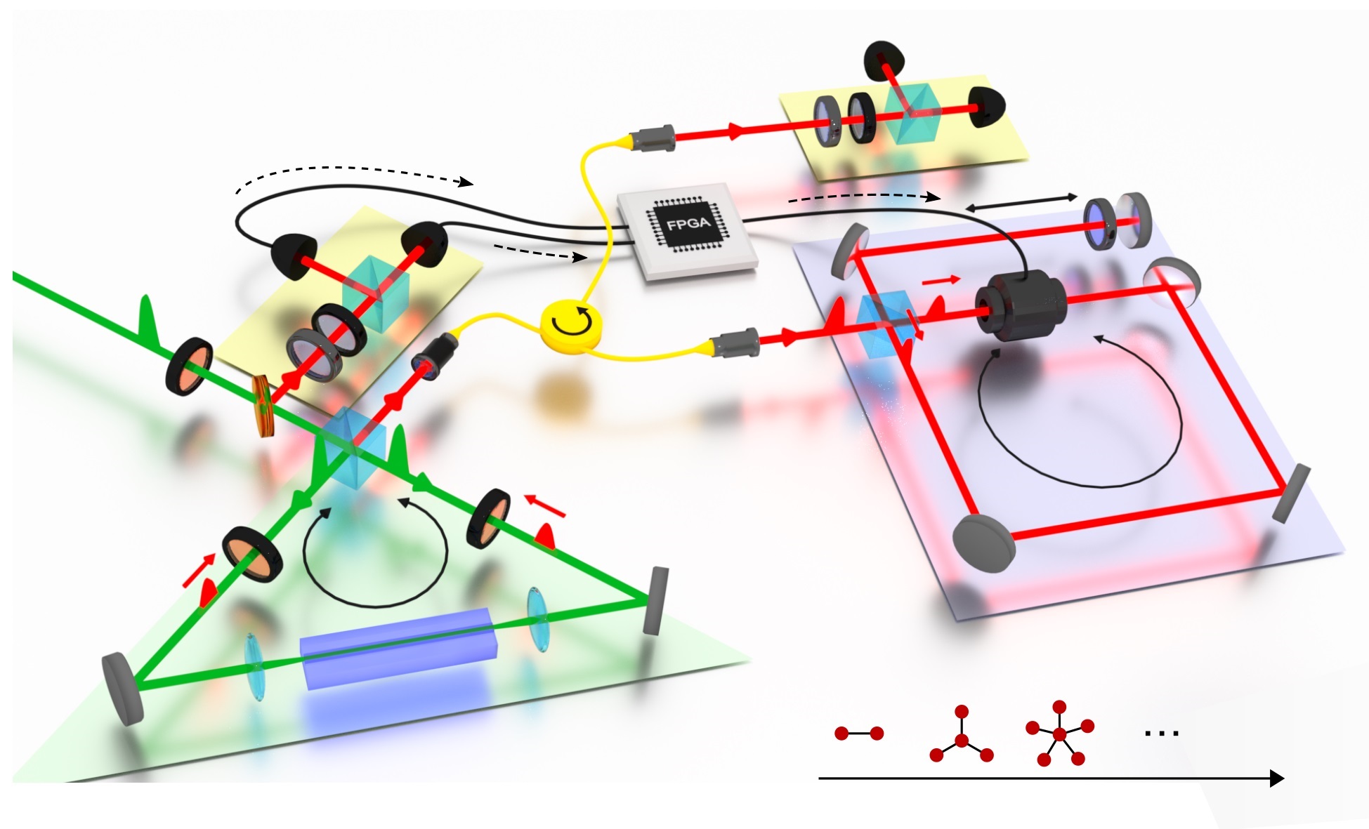
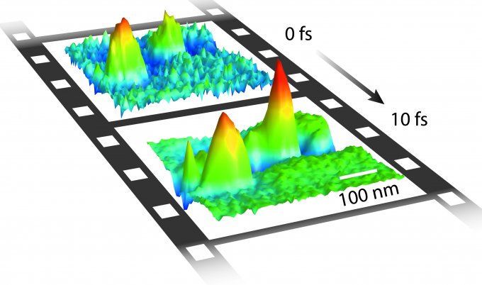
The ultrafast optical microscope assembled by the research team is 1,000 times more powerful than a conventional optical microscope, said CU-Boulder physics Professor Markus Raschke, lead study author. The “image frame” rate, or speed captured by the team, is 1 trillion times faster than the blink of an eye, allowing the researchers to make real-time, slow-motion movies of light interacting with electrons in nanomaterials - in this case a thin gold film.
"This is the first time anyone has been able to probe matter on its natural time and length scale," said Raschke. “We imaged and measured the motions of electrons in real space and time, and we were able to make it into a movie to help us better understand the fundamental physical processes."
A paper on the subject appears in the Feb. 8 issue of Nature Nanotechnology.
Matter is sometimes described as the “stuff of the universe” – the molecules, atoms and charged particles, or ions, that make up everything around us. Matter has several states, most prominently solid, liquid and gas.
According to the CU-Boulder researchers, a number of important processes like photosynthesis, energy conversion and use, and biological functions are based on the transfer of electrons and ions from molecule to molecule. The team used a technique called “plasmonic nanofocusing” to focus extraordinarily short laser pulses into tiny bits of gold film matter using a nanometer-sized metal tip.
“Our study brings nanoscale microscopy to the next level, with the ability to capture detailed images evolving on extremely fast time scales,” said Vasily Kravtsov, a CU-Boulder graduate student in physics and first author of the paper.
Other co-authors on the Nature Nanotechnology paper include CU-Boulder postdoctoral researcher Ronald Ulbricht and former CU-Boulder postdoctoral researcher Joanna Atkin, now a faculty member at the University of North Carolina-Chapel Hill.
“This work expands the reach of optical microscopes,” said Raschke. “Using this technique, researchers can image the elementary processes in materials ranging from battery electrodes to solar cells, helping to improve their efficiency and lifetime.”
Unlike electron microscope approaches, the new technique does not require ultra-high vacuum techniques and is particularly promising for studying ultrafast processes like charge and energy transport in soft matter, including biological materials, said Kravtsov.
The study was funded in part by the National Science Foundation and with support from the Pacific Northwest National Laboratory.