08/15/2016
By Edwin L. Aguirre
Scientists from UMass Lowell and King’s College London in the U.K. have demonstrated a new way of capturing ultrasharp images of structures of extremely tiny objects measuring billionths of a meter in size.
Called “interscale mixing microscopy,” or IMM, the technique can obtain details in viruses and nanoparticles much smaller than the wavelength of light. Such technology would be helpful in developing new vaccines against pathogens as well as innovative nanomaterials for industrial applications and novel pharmaceutical drugs to fight diseases.
“Our research addresses a fundamental problem in the field of microscopy,” says physics Prof. Viktor Podolskiy, who is the principal investigator for the UMass Lowell team. “When an object is smaller than the wavelength of light, you cannot really resolve the object’s size, shape or structure. Our technique is designed to go beyond this so-called ‘diffraction limit.’ ”
Podolskiy adds: “Sub-wavelength imaging with IMM can potentially be used to obtain the colors, or spectra, of small objects such as bacteria, viruses and nanoparticles. By knowing their color signatures we can rapidly identify and characterize the objects and determine their precise chemical composition.”
The team’s findings were recently published in Optica, the prestigious journal of The Optical Society. Funding for the research was provided by the U.S. National Science Foundation and the U.K.’s Engineering and Physical Sciences Research Council, Royal Society and Wolfson Foundation.
A Cost-effective Alternative to Electron Microscopes
“Conventional optical microscopes, such as those found in biology classrooms and hospital labs, use lenses to bend light and form images of everything, from tissues down to dust, pollen and blood cells,” explains Podolskiy. “However, objects whose size is smaller than the wavelength of light cannot be seen or even detected with these optical microscopes.”
He says while other imaging techniques, such as fluorescence microscopy, electron microscopy or scanning near-field microscopy, can in principle be used to assess the properties of small objects, none of these techniques is versatile or rapid enough in imaging and characterizing relatively large objects.
“A scanning electron microscope [SEM] has a tip that scans the surface of an object point by point. You then record the backscattering of light from that tip to build up an image,” says Podolskiy. “For large objects, this can take a long time.”
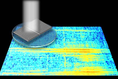
The IMM technique uses a conventional optical microscope and ingenious signal processing to decode the object’s properties based on the measurement of light that gets scattered by the object in close proximity to a special, finely ruled plate called a diffraction grating. The researchers showed that a single measurement with the grating may be enough to decipher with great precision the position, size and optical spectrum of the object.
SEMs can typically resolve details down to 5 to 10 nanometers. Right now, the IMM is constrained to about 70 nanometers.
“Although our technique is not yet as powerful, a brand-new scanning electron microscope can cost anywhere from hundreds of thousands of dollars to a million. The IMM can be retrofitted to older, existing research optical microscopes, thereby saving universities and companies a lot of money,” notes Christopher Roberts, a Ph.D. student in physics who conducted the project’s data processing and analysis.
He adds: “Moreover, you can’t observe living cells in an electron microscope; you have to kill and prepare them first. The IMM, in principle, can be used to observe live specimens in real time. Our technique could pave the way for the next generation of optical microscopy and nanoscale spectroscopy.”