July 21, 2015
By Leah Burrows
A new microscope capable of capturing nearly 17 billion pixels in a single image has been developed by Antony Orth PhD ’14, now of RMIT University in Melbourne, Australia, and Ethan Schonbrun of Harvard’s Rowland Institute. The microscope speeds up cellular imaging, making it easier to observe how cells respond to drug treatments. The research was published in the journal Optica.
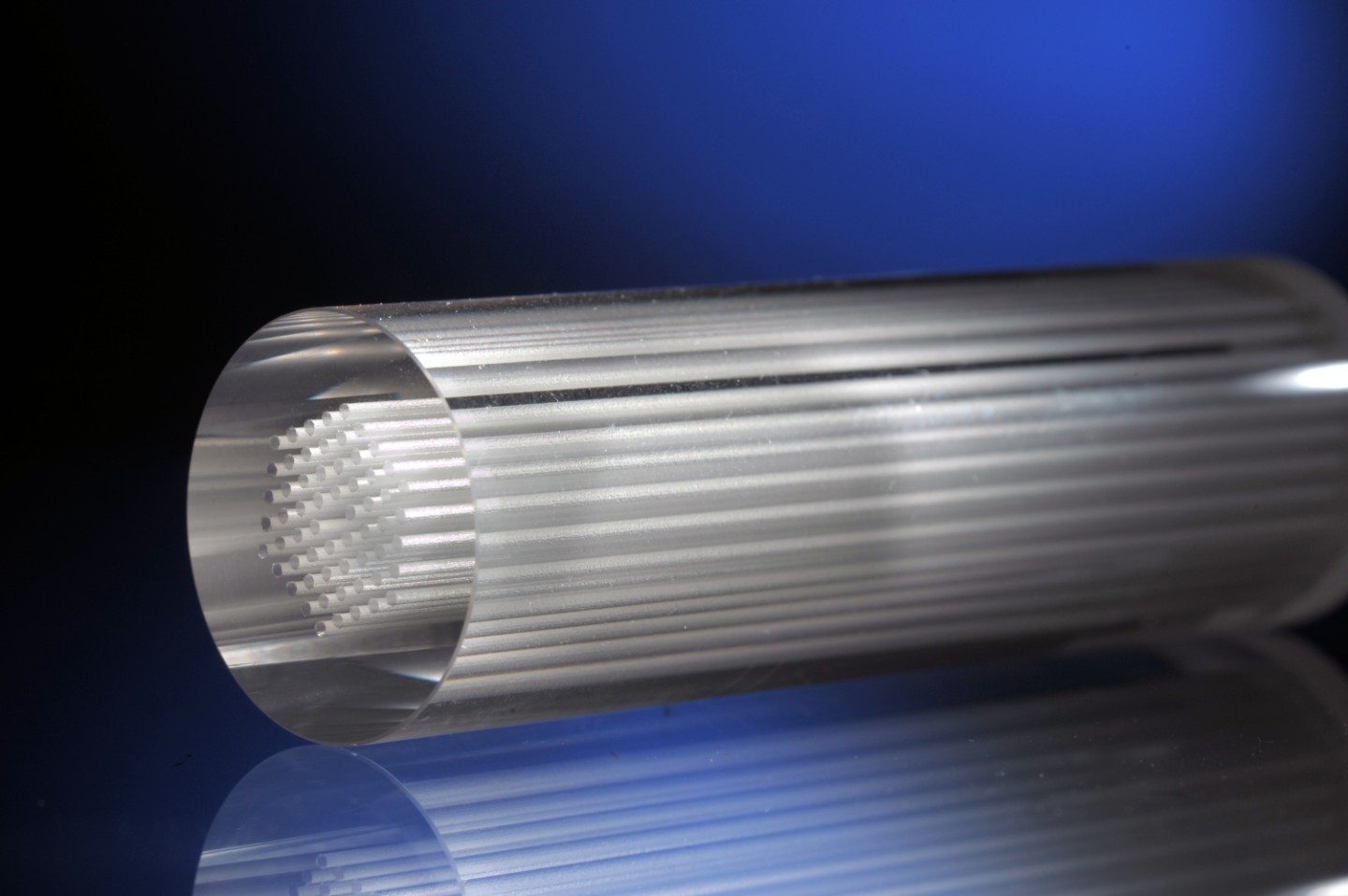
The microscope uses thousands of microlenses — each about the width of a human hair — and a dispersive prism to capture thousands of images and spectra simultaneously. The individual images are stitched together into one large mosaic, enabling the visualization of more than 13 resolvable colors in an extraordinarily large number of cells.
To zoom in, view the image on GigaPan.