02 December 2014
An opening in the mouse skull, or cranial window, enables imaging the neurons in the living brain using two-photon microscopy. But using this approach, one can generally only image the most superficial layers of the cortex. To overcome this, Andermann et al. use microprisms. They inserted 1-mm-sized glass prisms into the mouse brain, under the cranial window. The hypotenuse of the microprism is coated with aluminum and serves as a mirror that projects the incident light. In this way, the researchers imaged across the vertical plane of the prism and can view the brain in real time.
Microprisms Offer New Insights into Neural Networks
Thanks to techniques pioneered by Yale research scientist and lecturer Michael Levene and his team, the world may soon know more about how information is processed in the brain.
Levene's research focuses on the cortex, the outermost structure of the brain. As part of the brain's "grey matter," the cortex is responsible for aspects of memory, spatial reasoning, sensory perceptions, and even consciousness.
However, though proximity to the skull might make the cortex seem "accessible," imaging all six layers of the cortex has proven difficult. The most utilized in vivo imaging solution, two-photon microscopy, has trouble imaging the deeper cortical layers with high sensitivity and contrast because the tissue layers disperse the light. Moreover, this approach cannot image multiple layers simultaneously, limiting observation of interactions among the neurons in each layer.
To overcome these challenges, Levene's team implanted a glass microprism into the cortex of a mouse. One face of the prism was exposed along the brain's surface, while the other faces were inserted into the brain matter—one face perpendicular to the surface, another at 45-degree angle and coated with reflective aluminum. When a two-photon microscope was pointed at the exposed face, the microprism acted like a "microperiscope," enabling the researchers to simultaneously view all six cortical layers.
This latest study, published in Neuron, improves on Levene's previous microprism use to enable long-term implantation. Thus, while inserting the microprism did have some effect on the neurons nearest the insertion point, the structural integrity and health of most observed neurons was preserved. Neuron behavior could therefore be repeatedly imaged over a period of months, and, in fact, the clarity of the images progressively improved over time.
Levene's team—which included biomedical engineering postdoctoral fellows Nathan Gilfoy and Markus Wölfel, in addition to professor David McCormick from the Yale School of Medicine and Mark Andermann, Glenn Goldey and Clay Reid of Harvard Medical School—hope their methods will extend the reach of two-photon imaging. "Future integration with other optical approaches, like optogenetics, is going to be really exciting," says Levene. "We think this technique has a great future, and is going to make a big impact in neuroscience."
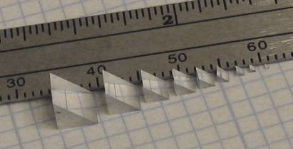
Tower Optical, your microprism source:
Tower Optical has been supplying microprisms to the medical industry since 2005 and is the ultimate source for optical components of all kinds. We have 9 standard sizes available: 0.5, 0.7, 1.0, 1.5, 2.0, 2.5, 3.0, 4.0, and 5.0 mm, available both coated and uncoated from stock.
During the ensuing years from introduction, Tower has made many custom variations for specific requirements.
This article was originally published on Tower Optical website. Read the original article.