29 APRIL 2013
To obtain very-high-resolution 3D images of the cerebral vascular system, a dye is used that fluoresces in the near infrared and can pass through the skin. The Lem-PHEA chromophore, a new product outclassing the best dyes, has been synthesized by a team from the Laboratoire de Chimie (CNRS / ENS de Lyon / Université Claude Bernard Lyon 1). Conducted in collaboration with researchers from the Institut des Neurosciences (Université Joseph Fourier - Grenoble / CEA / Inserm / CHU) and the Laboratoire Chimie et Interdisciplinarité: Synthèse, Analyse, Modélisation (CNRS / Université de Nantes), this work has been published online in the journal Chemical Science. It opens up significant prospects for better observing the brain and understanding how it works.
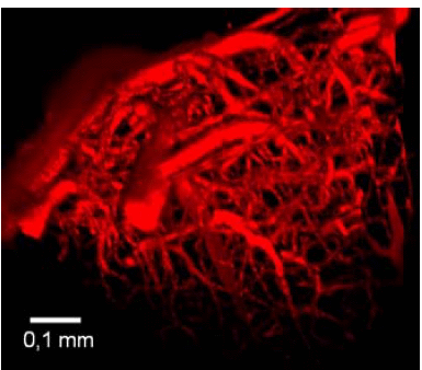
Different cerebral imaging techniques, such as two-photon microscopy or magnetic resonance imaging (MRI), contribute to our understanding of how the healthy or diseased brain works. One of their essential characteristics is their spatial resolution, in other words the dimension of the smallest details observable by each technique. Typically, for MRI, this resolution is limited to several millimeters, which does not make it possible to obtain images such as the one below, whose resolution is of the order of a micrometer.
To obtain such images of the vascular system of a mouse brain, it is necessary to use a fluorescent dye that combines several properties: luminescence in the near infrared, solubility in biological media, low cost, non-toxicity and suitable for 3D imaging (two-photon absorption). The researchers have developed a new product, Lem-PHEA, which combines these properties and is easy to synthesize. When injected into the blood vessels of a mouse, it has revealed details of the rodent's vascular system with previously unattained precision, thanks to a considerably enhanced fluorescence compared to “conventional” dyes (such as Rhodamine-B and cyanine derivatives). With Lem-PHEA, the researchers have obtained more contrasted images (in terms of brilliance) than with these standard dyes. Finally, the product is easily eliminated by the kidneys and no toxic residues have been found in the liver. These results pave the way for a better understanding of the working of the brain.
Bibliography
A water-soluble probe with near infra-red two-photon absorption and polarity-induced fluorescence for cerebral vascular imaging. Julien Massin, Azzam Charaf-Eddin, Florence Appaix, Yann Bretonnière, Denis Jacquemin, Boudewijn van der Sanden, Cyrille Monnereau and Chantal Andraud Chemical Science, first published on-line on the 22 April 2013.
DOI: 10.1039/C3SC22325F