07.03.2015
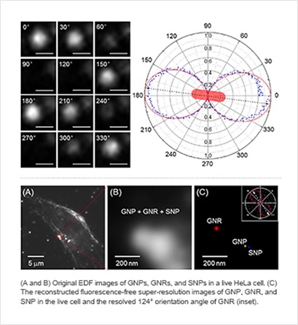
Professor Seongho Kang and his team at the Department of Applied Chemistry developed a fluorescence-free super-resolution optical microscopy technology that can for the first time in the world distinguish a distance as small as 2.5 nanometer (nm, 10-9 meters) between nanoparticles (NP) inside a living cell as separate entities. The research findings were published inScientific Reports, a Nature publication, on June 15, 2015. Professor Kang led the project; Peng Zhang (lead author) and Seungah Lee were the principal authors.
The laws of physics dictate that within the visible light wavelength the maximum theoretical resolution of conventional optical microscopy is limited by diffraction to roughly 200 nm. This limit, once deemed absolute, has now been overcome with the advent of super-resolution fluorescence optical microscopy technology, which improved the resolution down to about 20nm. This technology enabled unprecedented in vivo observation of sub-cellular structures, but was also fraught with such issues as high cost, low photostability, susceptibility to photobleaching, and carcinogenic risk from fluorescent materials employed.
In order to bridge these limitations, Professor Kang’s team has utilized plasmonic nanoparticles (NPs) instead of fluorescent materials in developing an enhanced dark-field (EDF) illumination technology based on wavelength-modulation that can resolve NPs inside cells less than 5nm apart. Professor Kang said, “We have now achieved a level of maturity for the base technology we developed. This super-resolution microscopy technology can have diverse applications in, for example, medicine, pharmacology, molecular biology, semiconductor, catalyst, surface treatment, and others.”
Direct collection of molecular information from DNA
Being able to observe in vivo sub-cellular micro structures through fluorescence-free super-resolution optical microscopy opens up many new possibilities in terms of studying the mechanism of pathogenesis, functions of nanoparticles in the cell cycle, scatter patterns of biomolecules, and others. Professor Kang is planning to research the ways to expedite diagnosis of infectious diseases by collecting genetic information directly from DNAs inside cells. In case of highly infectious viral diseases such as influenza and Middle East Respiratory Syndrome (MERS), for instance, the conventional diagnosis method using the polymerase chain reaction (PCR) requires time to amplify the DNA information and runs the risk of contamination. Professor Kang said, “If we can collect molecular information directly from DNAs inside the cell, it will greatly enhance the speed and accuracy of diagnosis for any disease.”
Translated by Tae Hun Kim