28 February 2014
The ability to locate and count small numbers of impurity atoms could lead to advances in modern electronics and optical fiber communication networks.
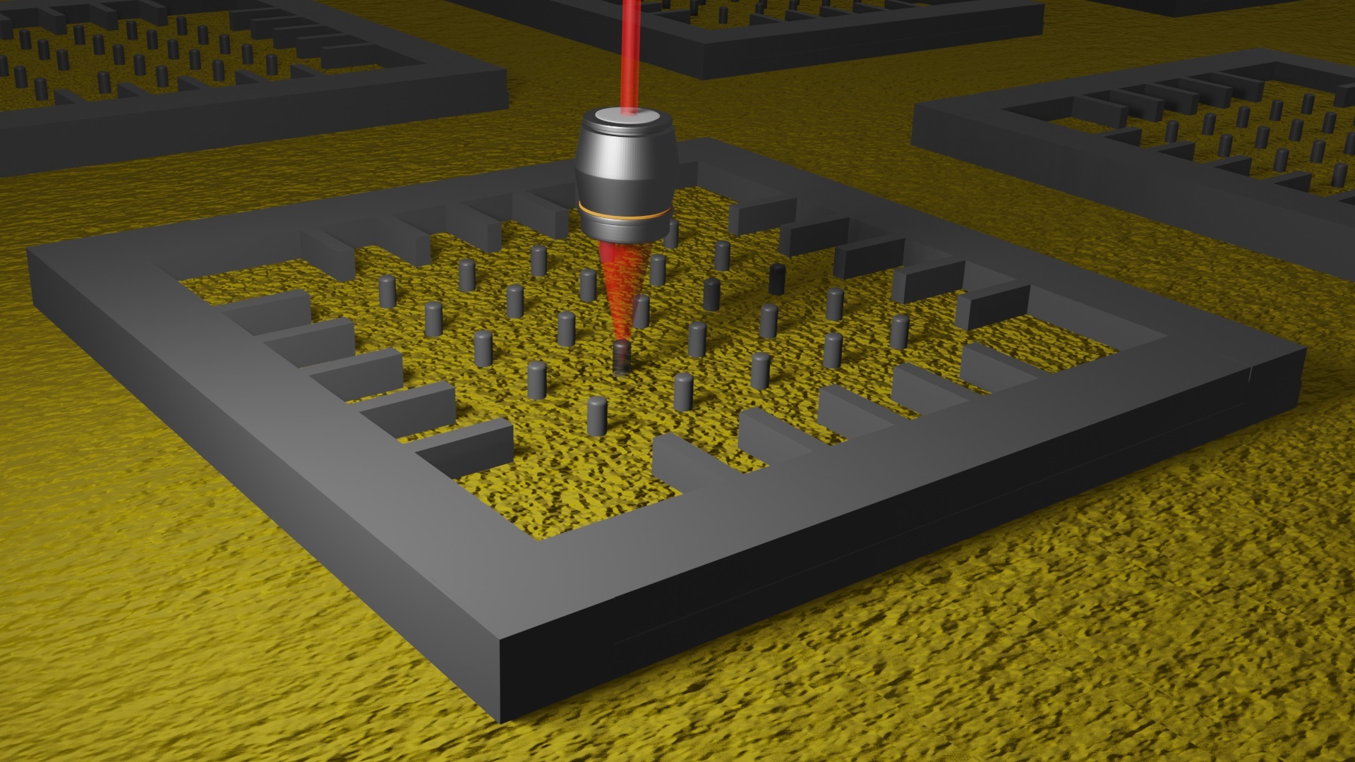
In research published today in Physical Review Letters, physicists from Monash University, the University of Melbourne and TU Graz, Austria, show a method called spectrum imaging can be used to measure atom concentrations at atomic resolution.
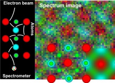
By using spectrum images to visualise where atoms are and how they are bonded, scientists will gain further insight into the properties of new materials. Spectrum imaging provides a digital image encoding this complex information through colour.
Co-author Dr Scott Findlay, of Monash University’s School of Physics, said the technique could be a useful tool to characterise new materials.
“When probed with an electron beam, atoms give that beam an energy spectrum in a way that is like adding colour. Distinct atomic species add distinctive colours,” Dr Findlay said.
“Imagine putting several open paint tins inside a waterfall. With careful measurements on the pool below, one could determine not just the colours but also the number of different tins used. Type and number – that’s quantitative spectrum imaging at low spatial resolution.”
However, precision analysis at atomic resolution is more challenging.
“Spectrum imaging at atomic resolution is more like a game of pinball with different coloured, freshly-painted pins,” Dr Findlay said.
“If the ball ends up with five red, two blue and one green paint-spots, that doesn’t necessarily tell us the number of differently coloured pegs present, just those that were hit on the path the ball happened to take.”
Dr Findlay said scanning an electron beam across the sample allowed them to map the specimen structure but also provided the information needed to untangle the problem.
“With an understanding of how the electron beam interacts with the specimen – how the ball bounces – we can establish both the location of the atoms – the pins – and their species – the colours,” Dr Findlay said.
“The ability to map out the concentrations of different atomic species at atomic resolution is a significant step towards developing new materials and technology.”
Dr Findlay said more research was needed to develop the new technique further.
Dr Findlay, with Associate Professor Matthew Weyland of the Monash Centre for Electron Microscopy and colleagues at the University of Melbourne, has been awarded an Australian Research Council Discovery Project grant to continue their work.