July 22, 2013
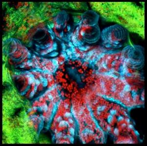
Corals are beautiful when seen through your own eyes in sunlight, but for scientists at the University of Hawaiʻi at Mānoa, seeing corals in this manner is simply not enough.
As part of a new PBS digital video series called UnderH20, viewers see live corals--and their associated microorganisms--from a whole new perspective. Using a revolutionary new tool, a laser scanning confocal microscope, scientists at the Hawaiʻi Institute of Marine Biology generate images that are one part art and one part science.
In this video, they take us along for a ride with this amazing new technology and show us corals in a way that we have never seen before.
The laser scanning confocal microscope at HIMB that visualizes the beautiful biology captured in this video was purchased with funds donated by Pam Omidyar in 2010.
For more information about the coral reef research related to the visualization, visit:
http://www2.Hawaiʻi.edu/~rgates/Gates_Lab_Website/Gates_Lab.html http://www.Hawaiʻi.edu/himb/facilities/confocal.html
For more information, visit: http://www.hawaii.edu/himb/