November 2, 2017
Scientists gain an insight into the fascinating world of atoms and molecules using x-ray microscopes. Ground-breaking research by physicists at FAU, the Deutsches Elektronen Synchrotron (DESY) in Hamburg, and the University of Hamburg has paved the way towards new imaging techniques. The team of scientists have successfully developed and tested a method which is considerably more effective than conventional procedures. With conventional imaging methods using the diffraction of coherent light, scientists have to go to considerable lengths to ensure the coherence of the radiation, i.e. the electromagnetic waves must remain in phase during the scattering process. The new method uses incoherent light instead. The process, which has now been demonstrated for the first time using short-wave ultraviolet radiation, stands to revolutionise the methods of diffractive optics and has been published in the journal Nature Physics.
Conventional methods researchers use to determine the structure of crystals and minerals are based on the coherent scattering of light. In other words, light waves hit a structure and are deflected, but continue to oscillate without their pattern of crests and troughs being distorted or interrupted in any way. If a sufficient number of these photons can be measured with a detector, a characteristic diffraction pattern is obtained which can be used to derive the pattern of scattered atoms or the crystal structure.
Most light waves, however, are scattered incoherently, that is the wave patterns of the outgoing waves are no longer directly in relation to the incoming waves as the light is reflected from the atoms it touches as fluorescent light. The result is diffuse background light which scientists have until now believed was not suitable for imaging, having a negative effect on the accuracy of the method.
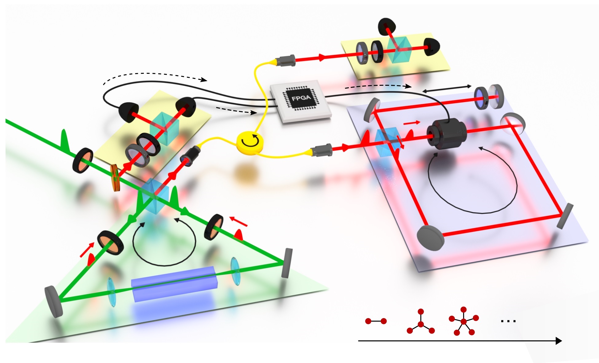
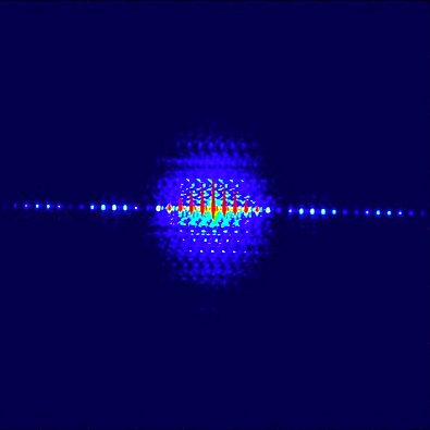
Now, scientists have used the very same incoherent radiation to record a structure shaped like a benzene ring with the help of diffuse scattered light in the soft X-ray range at DESY’s free-electron laser FLASH. “Our work provides the basis for a fundamentally new approach to X-ray imaging, because our experiment has demonstrated that incoherently scattered photons can be used to reconstruct complex structures at short wavelengths,” explains Raimund Schneider of FAU, the principal author of the paper on the experimental findings.
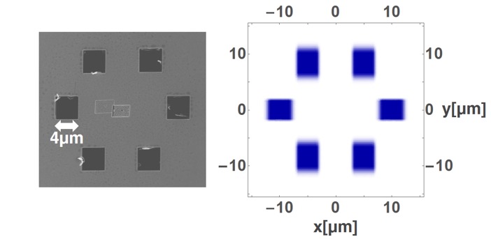
Image of investigated hexagonal benzene ring structure taken with an electron microscope. Right: Reconstructed image of the benzene ring structure taken using the innovative method based on incoherent light. In future, this method could be used to obtain images of individual atoms with an even higher resolution using short wave x-rays.
Image: Nature Physics
This incoherently scattered light, however, is precisely what has now been used to analyse a structure. At DESY, the researchers successfully created an image of a hexagonal, micrometre sized structure in the shape of a benzene ring. The basic technique behind this procedure is not new. Robert Hanbury Brown and Richard Q. Twiss used incoherent light to determine the diameter of stars as early on as 1956. The team of researchers from Erlangen and Hamburg have now refined this method, using it to analyse microscopic structures.
The innovative method has one decisive advantage. ‘The smaller the structures to be imaged, the larger the proportion of incoherently scattered light,’ explains the lead author of the study, Raimund Schneider from FAU. ‘Whilst this poses coherent imaging increasing problems with intensity, our method actually benefits from it.’ The new method has the potential to achieve a significant improvement in analysing structures in the fields of biology and medicine.
The original publication ‘Quantum Imaging with incoherently scattered light from a free-electron laser’ was published in Nature Physics.
Reference: >„Quantum Imaging with incoherently scattered light from a free-electron laser“; Raimund Schneider, Thomas Mehringer, Giuseppe Mercurio, Lukas Wenthaus, Anton Classen, Günter Brenner, Oleg Gorobtsov, Adrian Benz, Daniel Bhatti, Lars Bocklage, Birgit Fischer, Sergey Lazarev, Yuri Obukhov, Kai Schlage, Petr Skopintsev, Jochen Wagner, Felix Waldmann, Svenja Willing, Ivan Zaluzhnyy, Wilfried Wurth, Ivan A. Vartanyants, Ralf Röhlsberger, Joachim von Zanthier; Nature Physics, 2017; DOI: 10.1038/nphys4301