29 January 2016
Super-sharp images from within the human body made through tiny endoscopes have come a step closer to reality thanks to joint research by scientists from the University of Twente's MESA+ research institute, the Max Planck Institute for the Science of Light (MPL), FOM and Carl Zeiss AG. An advanced wavefront shaping method developed at the UT combined with unique optical fibres from MPL make it possible to focus lensless light at an unparalleled resolution. FOM postdoc Dr Lyubov Amitonova and her colleagues published their findings on 29 January in Optics Letters, the leading journal of the Optical Society of America.
Optical imaging via ultrathin fibre probes is extremely useful for taking a look inside the human body in a minimally invasive manner. Unfortunately, the resolution of current fibre endoscopes is one micrometre at best. This is not enough to see interesting and important fine features in biological cells, for example. Some endoscopes use a high number of separate fibres bound together into a fibre bundle. Each fibre then acts like a discrete pixel to form the final pixelated image. However such bundles tend to be quite thick, at least a millimetre in diameter.
An alternative is fibre endoscopes based on 'multimode' fibres. These could offer imaging with a better view and be as thin as a tenth of a millimetre. A multimode fibre uses only a single fibre core that can transmit an entire image. Unfortunately, the image becomes scrambled as it passes through the fibre. However, some tricks for unscrambling these images are available. The main limiting factor for the resolution of these multimode endoscopes is that the fibres only transmit light that propagates along the fibre axis. Light under a small angle can still bounce from the fibre walls. But if this angle gets too large, the light will simply leak out to the side. FOM postdoc Dr Lyubov Amitonova and her colleagues at UT and MPL have now shown that with photonic crystal fibres this limitation can be overcome.
Unique crystal fibre probe
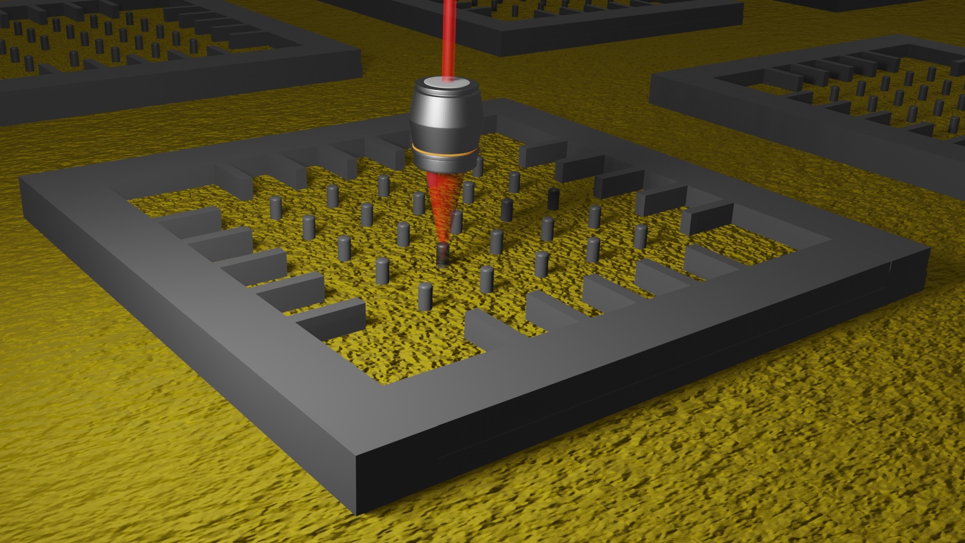
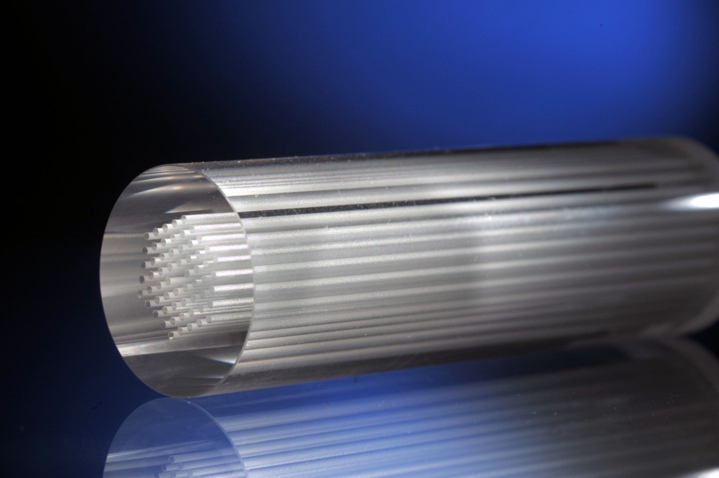
Conventional ('step-index') fibres consist of two zones of different material (an outer cladding and an inner core) with distinct refraction indices, which enable light transmission down the fibre axis by total internal reflection. Photonic crystal fibres are built differently: they are made of one material only and light guiding is realised through the presence of a specific pattern of holes in the cladding, which are filled with air. Tailoring the cladding structure of such a fibre provides a unique tool for engineering specific fibre-optic properties. In this project, the scientists have designed and made such a fibre to focus a laser beam through the fibre down to 0.52 micrometres, using visible red light.
Sharp focus and high resolution
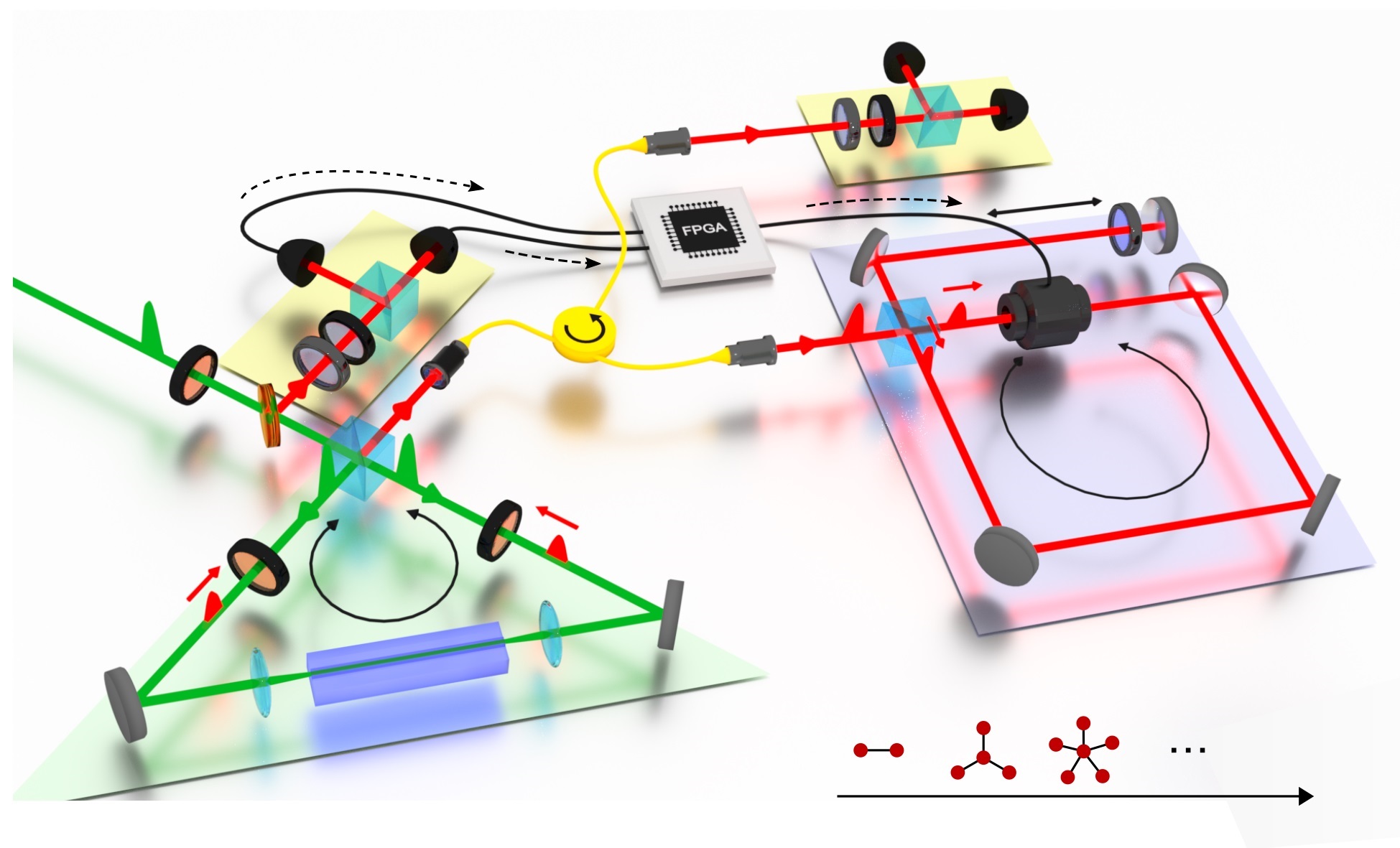
A photonic crystal fibre acts as a multimode fibre in which the image typically gets scrambled due to light bouncing off the possibly irregular wall of the fibre. The technique of complex wavefront shaping, invented at UT, is able to undo such scrambling and make a sharp focus. This is achieved by pre-shaping the light into the precise form needed to make a sharp image behind the fibre before the light actually enters the fibre. Using this approach, Amitonova and her colleagues have succeeded in focusing light at the fibre output facet of different multimode fibres including several unique photonic crystal fibres. They have shown that the complex wavefront shaping technique together with a properly designed multimode photonic crystal fibre allows the creation of a tightly focused spot at the desired position on the fibre output facet with a subwavelength beam waist. This paves the way towards high-resolution endoscopic imaging via fibre probes so thin that they could be inserted, for instance, into tiny blood vessels not much thicker than a human hair.
This research has been made possible by funding from the Foundation for Fundamental Research on Matter (FOM), Technology Foundation STW, and the Netherlands Organisation for Scientific Research (NWO).
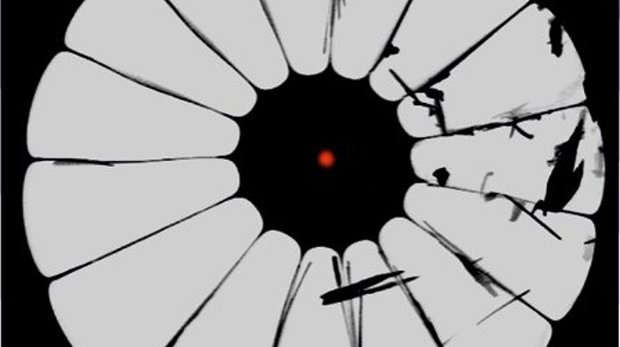
Illustration: Electron microscope image of one of the photonic crystal fibres made at MPL for this research. The glass is black, the air is grey and the red dot illustrates the focus.
Full journal reference: Lyubov V. Amitonova, Adrien Descloux, Joerg Petschulat, Michael H. Frosz, Goran Ahmed, Fehim Babic, Xin Jiang, Allard P. Mosk, Philip St.J. Russell, and Pepijn W.H. Pinkse, “High-resolution wavefront shaping with a photonic crystal fiber for multimode fiber imaging” Optics Letters 41, (2016).