30 May 2014
University scientists have developed a rapid new technique involving X-ray imaging that allows clear images to be obtained displaying the orientational properties of molecules in solid materials.
Published today in the journal Science, the novel technique which scientists are calling X-ray Birefringence Imaging (XBI) yields a map indicating the specific orientations of molecules in different regions of a material. The information garnered from this method will be of benefit to scientists in a variety of fields in revealing detailed knowledge of the orientational characteristics of individual molecules and bonds within materials.
Developed by researchers from Cardiff University and Diamond Light Source, the principles underlying the X-ray Birefringence Imaging technique follow a close analogy in concept to the polarizing optical microscope, used extensively across the physical, geological and biological sciences. However, rather than polarized visible light, the new X-ray Birefringence Imaging technique uses polarized X-rays to interrogate the material of interest, yielding an image of X-ray Birefringence across the whole material.
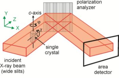
Professor Kenneth Harris from the Cardiff School of Chemistry, who led the research project, said: "Our paper is the first report of an experimental set-up that enables X-ray Birefringence measurements to be carried out in a spatially resolved manner, producing a direct image of the X-ray birefringence from all parts of the material under investigation."
The application of the X-ray Birefringence Imaging technique is demonstrated by experiments carried out under conditions that selectively detected the orientations of C–Br bonds in brominated organic solids, highlighting the utility and sensitivity of X-ray Birefringence for imaging the local orientational properties of materials, including characterization of changes in molecular orientational ordering associated with solid-state phase transitions.
Findings highlighted in the paper also show that X-ray Birefringence Imaging is a powerful tool for identification and characterization of "domain structures" in materials, in which different regions of the material comprise orientationally distinct arrangements of the component molecules, leading to an understanding of the size, spatial distribution and temperature-dependence of the domain structures.
Although the new method is demonstrated using single-crystal samples, there is no requirement for crystallinity, as X-ray Birefringence is sensitive specifically to local molecular orientations. Therefore, X-ray Birefringence Imaging could be applied to any material - including liquid or amorphous phases - with an anisotropic distribution of molecular orientations.
The X-ray Birefringence Imaging technique also has particular utility in cases (e.g. partially ordered materials, multiply twinned crystals, or other materials with complex domain structures) for which the application of diffraction-based techniques is either not straightforward or not feasible.
In addition to providing new insights on the structural anisotropy of a wide range of solid materials, future opportunities for applications of X-ray Birefringence Imaging include studies of liquid crystalline materials and oriented molecules at interfaces. Furthermore, as highlighted in theScience paper, X-ray Birefringence images can be recorded rapidly, leading to very exciting prospects for exploiting the time-resolved capabilities of X-ray Birefringence Imaging. Such applications would allowtime-dependent changes in molecular orientational distributions to be probed.
More information:
School of Chemistry